The POCUS Atlas
Point-of-care ultrasound, loop by loop
The POCUS Atlas is a peer-reviewed, open collection of exemplary point-of-care ultrasound clips contributed by clinicians worldwide. Every clip in its image galleries is here as a case: watch the loop, commit to the finding, then read the contributor's own description.
- Loops
- 1324 loops
- Galleries
- 19 galleries
- Normals
- 166 normal studies
- Reviews
- 56 one-minute reviews
Adult galleries
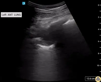
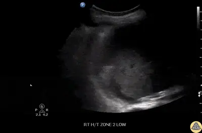
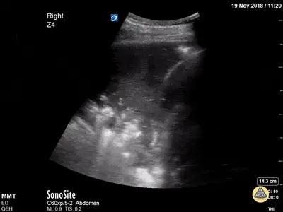
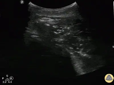
Lung
A-lines, B-lines, sliding, consolidation, effusion and the lung point.
Open the gallery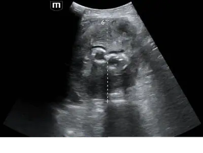
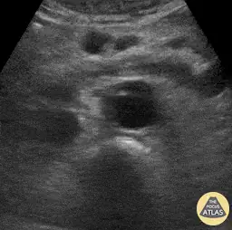
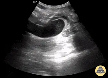
Aorta
Aneurysm, dissection flap, rupture and the normal calibre from the diaphragm to the bifurcation.
Open the gallery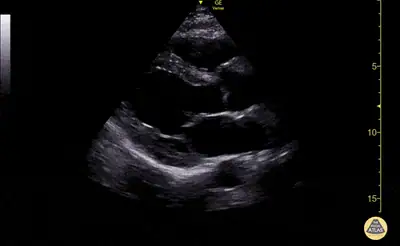
Echo anatomy
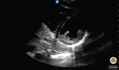
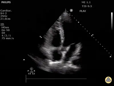
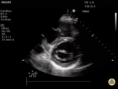
The standard windows, labelled and coloured: what every chamber looks like when nothing is wrong.
Open the gallery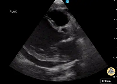
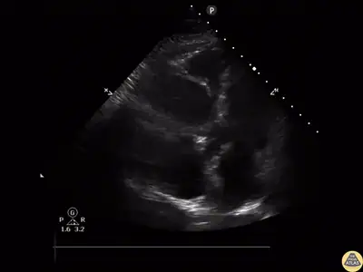
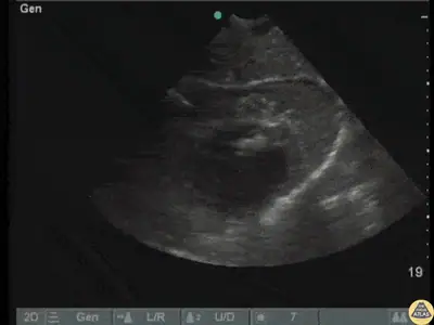
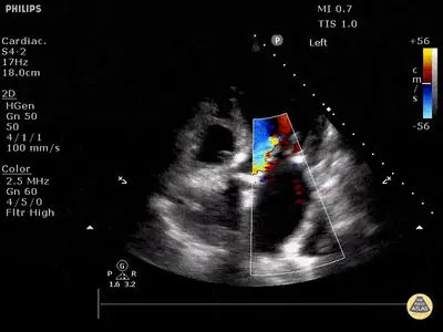
Echo
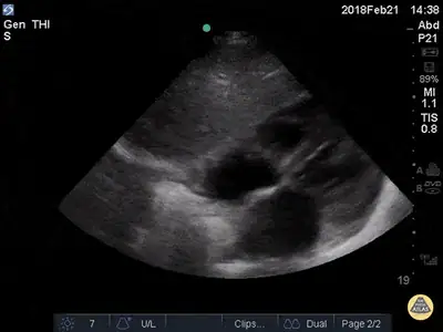
Ventricular failure, pericardial disease, valves, the IVC, masses and the unusual.
Open the gallery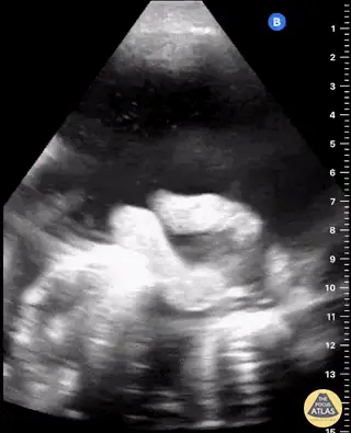
Bowel
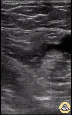
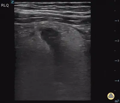
Obstruction, appendicitis, free fluid, hernia and the peristalsis that tells you what the bowel is doing.
Open the gallery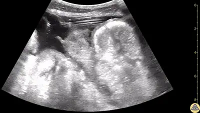
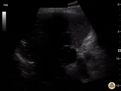
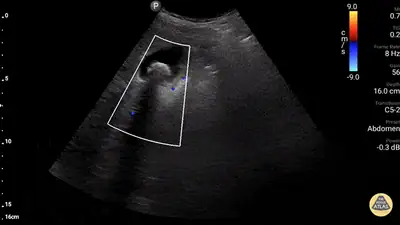
Hepatobiliary
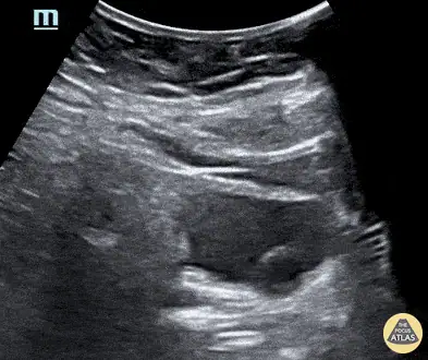
Gallstones, cholecystitis, the dilated duct, the liver and its vessels.
Open the gallery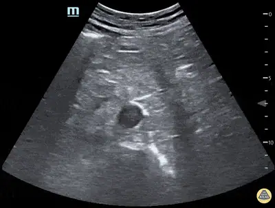
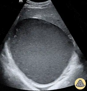
Renal and bladder
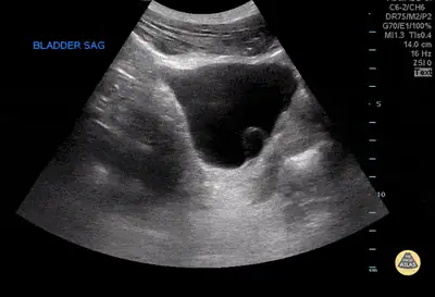
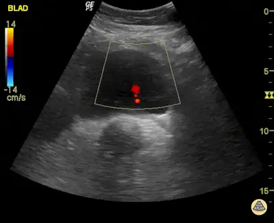
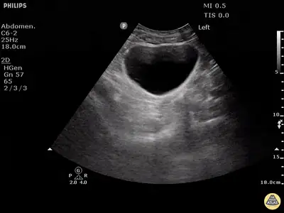
Hydronephrosis in every grade, stones, cysts, the bladder and the ureteric jet.
Open the gallery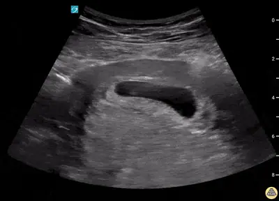
OB and gynaecology
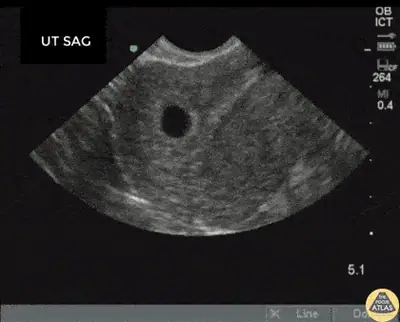
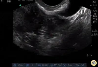
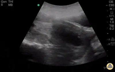
Intrauterine pregnancy, the ectopic, fetal heart activity, the ovary and free fluid.
Open the gallery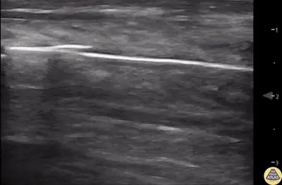
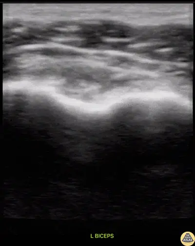
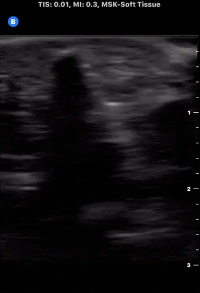
MSK and soft tissue
Abscess versus cellulitis, tendons, fractures, effusions, foreign bodies and the dislocated joint.
Open the gallery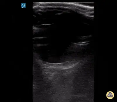
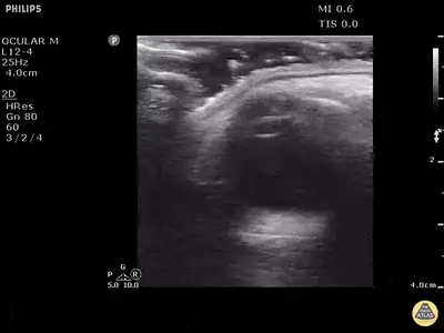
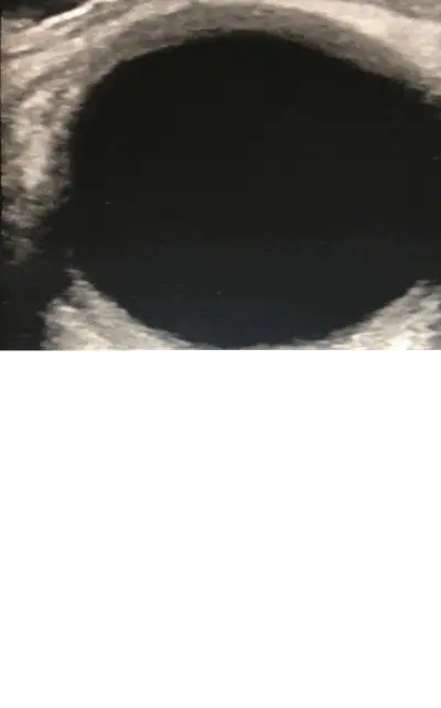
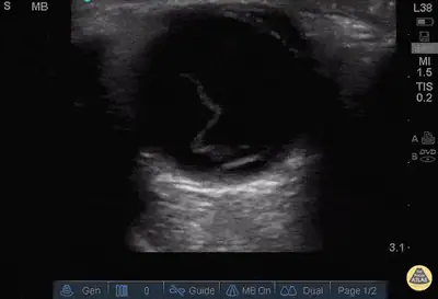
Ocular
Retinal and vitreous detachment, haemorrhage, the lens, the optic nerve sheath and the pupil.
Open the gallery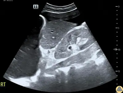
Trauma (FAST)
The FAST and eFAST windows: free fluid, haemothorax, pneumothorax and the pericardium after injury.
Open the gallery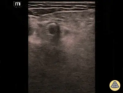
Vascular
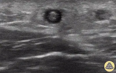
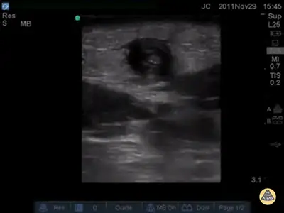
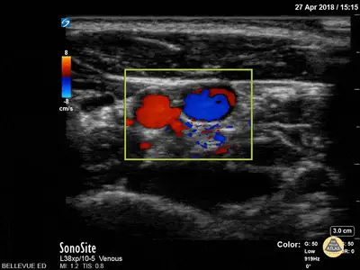
Compressibility, the clot that will not squash, superficial thrombosis, arterial pathology and access anatomy.
Open the galleryPOCUS Atlas Jr: paediatric galleries
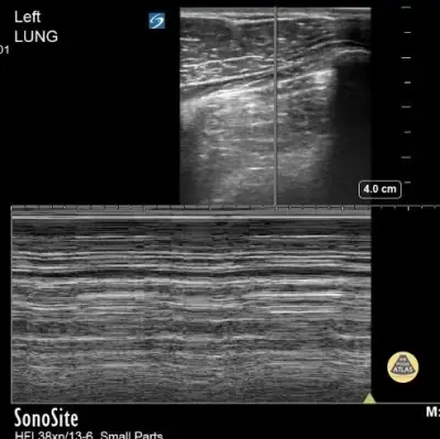
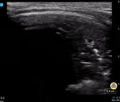
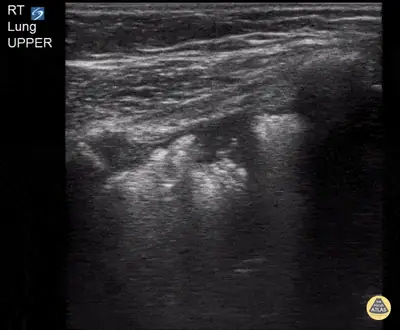
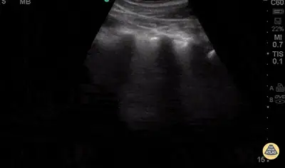
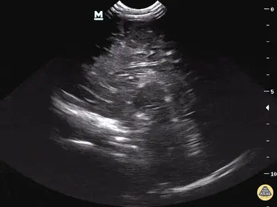
Paediatric lung
The child's chest on ultrasound: consolidation, bronchiolitis patterns, effusion.
Open the gallery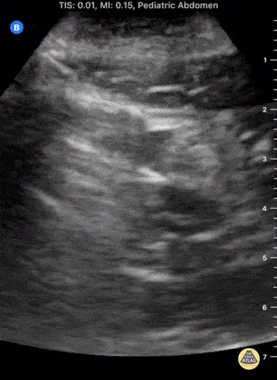
Paediatric GI
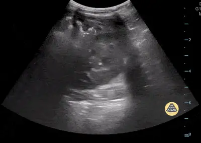
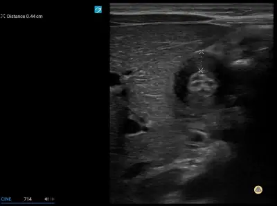
Intussusception, pyloric stenosis, appendicitis and the child's abdomen at the bedside.
Open the gallery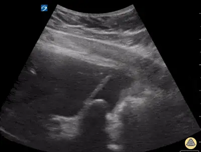
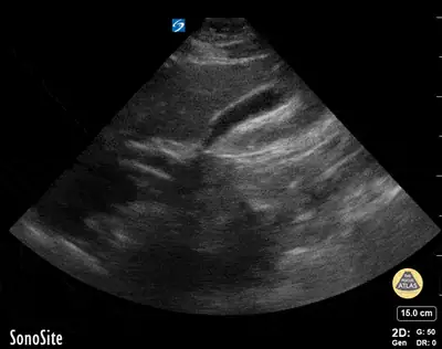
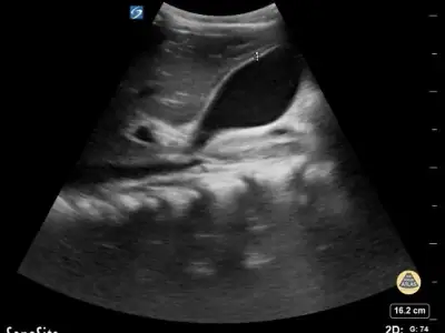
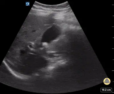
Paediatric biliary
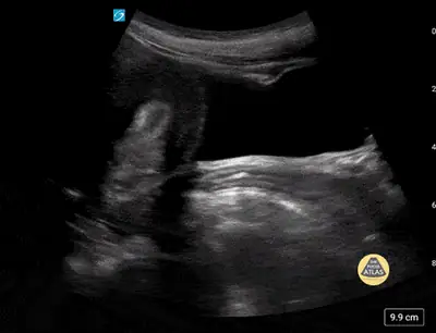
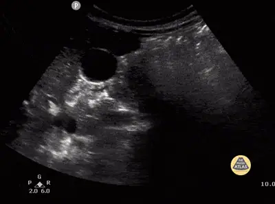
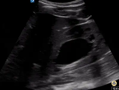
The child's gallbladder and liver at the bedside.
Open the gallery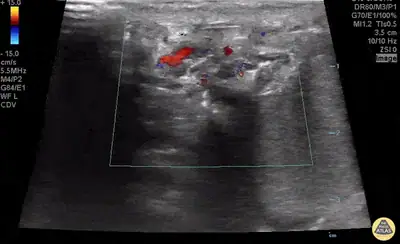
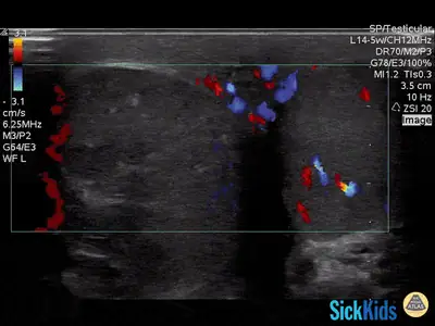
Paediatric GU
The child's kidney, bladder and scrotum on ultrasound.
Open the gallery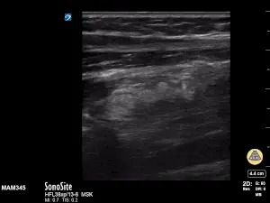
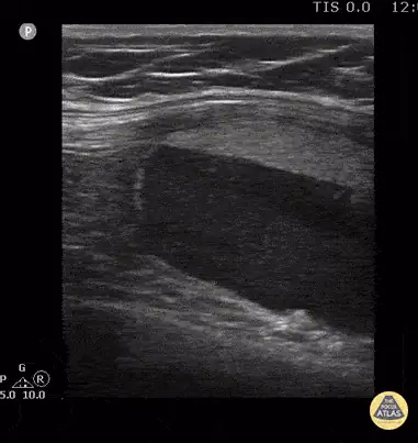
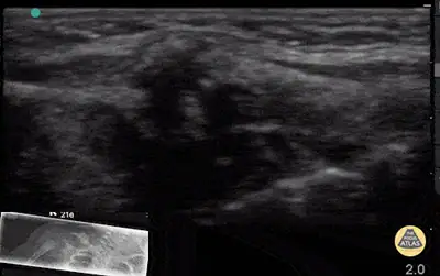
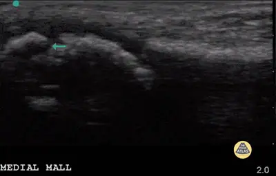
Paediatric MSK
Hip effusion, fractures, soft-tissue infection and the child's joint.
Open the gallery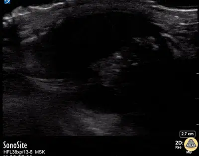
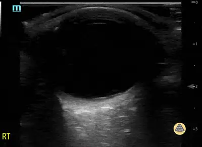
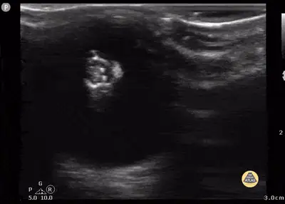
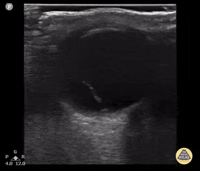
Paediatric orbit
The child's eye and orbit at the bedside.
Open the gallery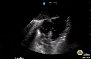
Paediatrics
The POCUS Atlas Jr mixed gallery: lung, abdomen, heart and more in children.
Open the gallerySource, licence and honesty
Clips and captions are from The POCUS Atlas, licensed CC BY-NC 4.0. Each case names its contributor where the Atlas does, links to the clip's own page, and carries the non-commercial flag: this material is for free education and must be reviewed before any commercial use.
The diagnosis on each case is the contributors' label as published by the Atlas. RadSpace has not separately reviewed these loops, so there is no click-to-locate step: you name the finding on the whole clip and are scored on the answer and your calibration.