Recognizing pneumothorax
Learning objectives
- Identify the visceral pleural line
- Distinguish pneumothorax from mimics
- Recognize features of tension
Step-by-step approach
- 1
The two-part definition
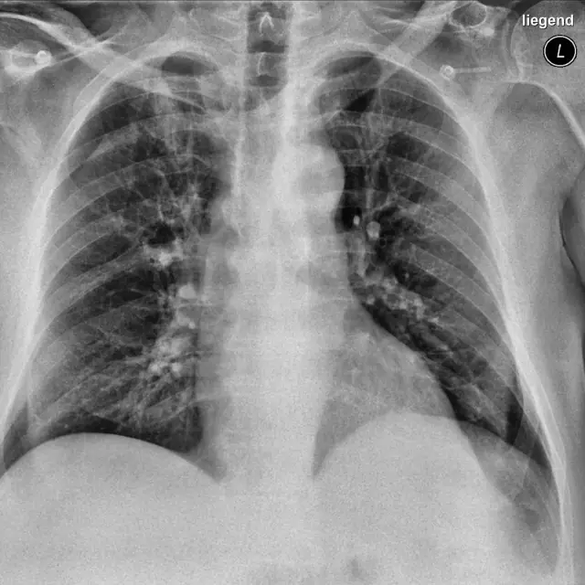
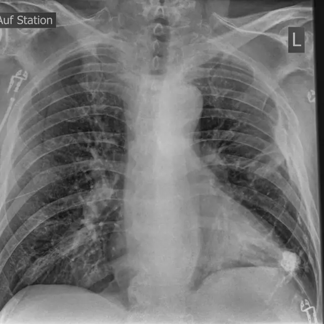
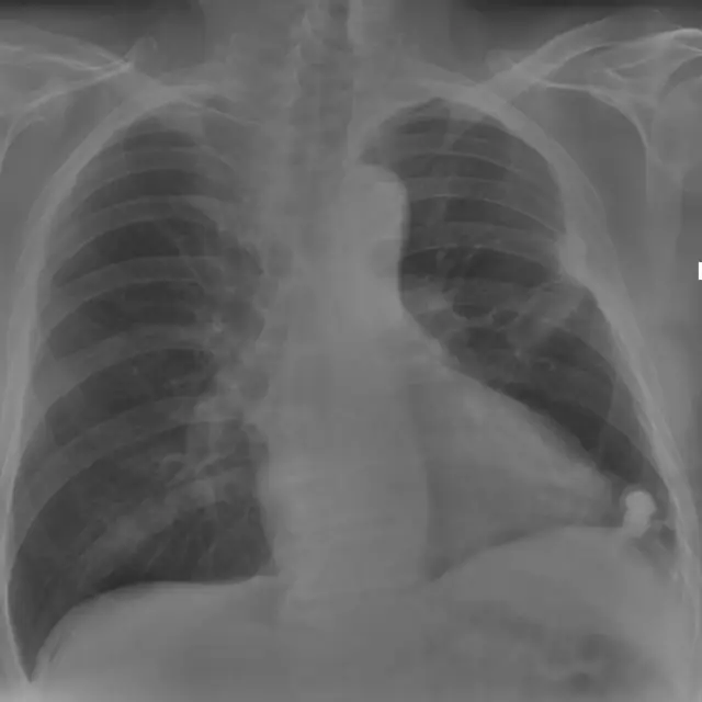
A pneumothorax on a radiograph is a line plus an absence: a thin visceral pleural line paralleling the chest wall, with no lung markings peripheral to it.
- 2
Where to look
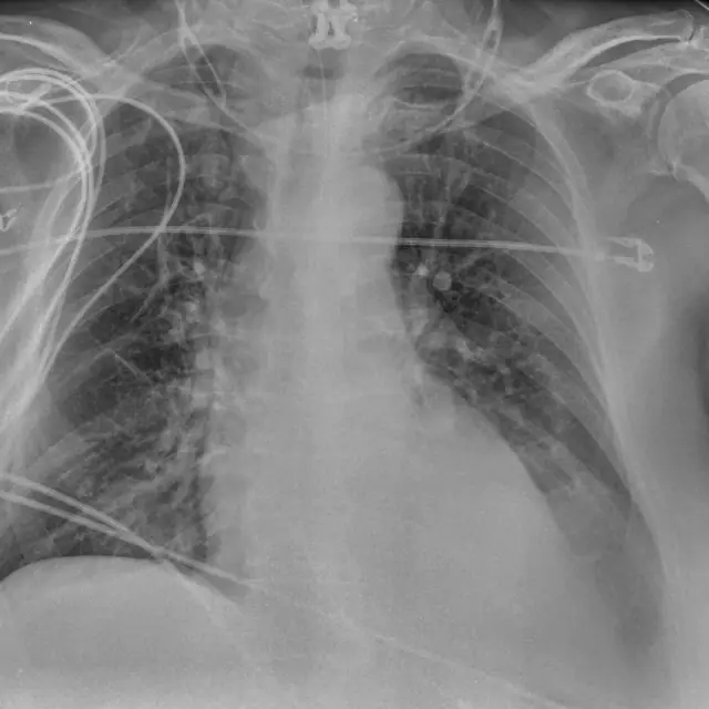
On erect films air rises to the apex, trace each pleural edge from the apex down. On supine films air collects anteriorly: look for the deep sulcus sign.
- 3
Mimics
Skin folds create edges (density change) rather than lines, and markings continue beyond them. Bullae have concave inner margins. The scapular edge follows the scapula.
- 4
Tension
Contralateral mediastinal shift, depressed hemidiaphragm, splayed ribs. This is a clinical emergency, the report should never delay decompression.
- 5
On ultrasound
Absent lung sliding with A-lines suggests pneumothorax; the lung point confirms it. Any B-line excludes it at that interspace.
Common mistakes
- Calling skin folds pneumothorax
- Missing the deep sulcus sign on supine films
- Waiting for imaging in clinically obvious tension
Summary checklist
- Pleural line present?
- Markings absent beyond it?
- Mediastinum central?
- Supine? Check the deep sulcus
Ask RADSpace about this guide
AI responses are for education and may contain errors. RADSpace is not for primary clinical diagnosis.