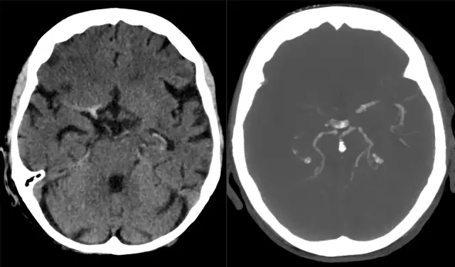
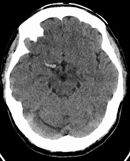
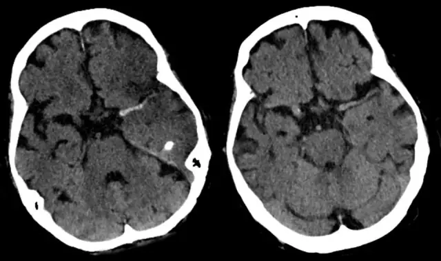
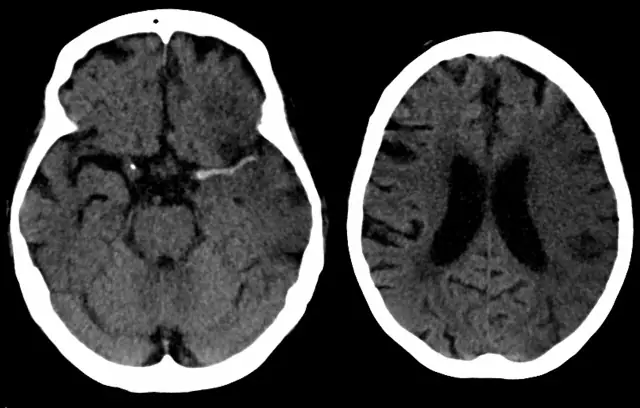
Recognizing intracranial hemorrhage
Learning objectives
- Differentiate the five compartments of intracranial blood
- Track how blood density evolves with time
- Report the modifiers that change management
Step-by-step approach
- 1
Density and time
Hyperacute-to-acute blood is bright; over 1–2 weeks it becomes isodense to brain, then hypodense. A subacute isodense subdural is a classic trap.
- 2
Extradural
Lentiform (lens-shaped), limited by sutures, usually arterial and post-traumatic, often with an overlying fracture.
- 3
Subdural
Crescentic, crosses sutures but not dural reflections; common in older patients and may follow trivial trauma.
- 4
Subarachnoid
Blood in sulci and cisterns. Non-traumatic SAH means aneurysm until proven otherwise.
- 5
Intraparenchymal and intraventricular
Location suggests cause: basal ganglia favours hypertension; lobar in older patients suggests amyloid; always note intraventricular extension.
- 6
The four modifiers
Every hemorrhage report needs: location, size, intraventricular extension, and midline shift/mass effect.
Common mistakes
- Missing isodense subacute subdurals
- Not switching windows to separate blood from adjacent skull
- Omitting mass-effect assessment
Summary checklist
- Which compartment?
- Acute or evolving?
- IVH?
- Midline shift?
- Cisterns open?
Ask RADSpace about this guide
AI responses are for education and may contain errors. RADSpace is not for primary clinical diagnosis.