Understanding common radiology terminology
Learning objectives
- Translate density/intensity/echogenicity terms across modalities
- Use localizing signs correctly
- Avoid the most common terminology errors
Step-by-step approach
- 1
Radiographs and CT: density
Lucent = darker (air); opaque/dense = whiter (bone, metal, contrast). On CT use hypo-/iso-/hyperdense, quantified in Hounsfield units: air −1000, fat ~−100, water 0, acute blood 50–70, bone 1000.
- 2
MRI: intensity
Hypo-/hyperintense, always with the sequence named: 'T2-hyperintense'. Fluid is bright on T2, dark on T1.
- 3
Ultrasound: echogenicity
Anechoic (black, fluid), hypoechoic, hyperechoic (bright). Shadowing and enhancement are artifacts that carry diagnostic meaning.
- 4
Localizing language
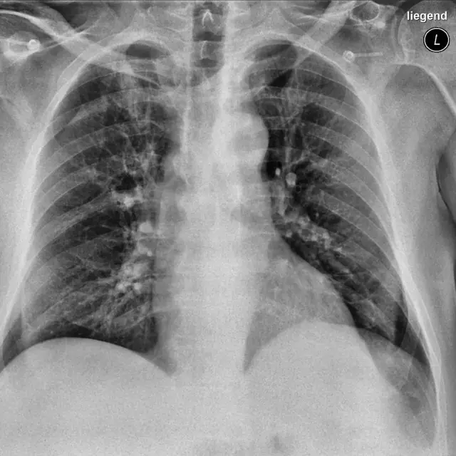
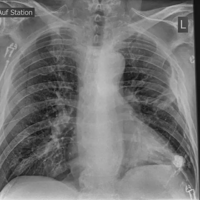
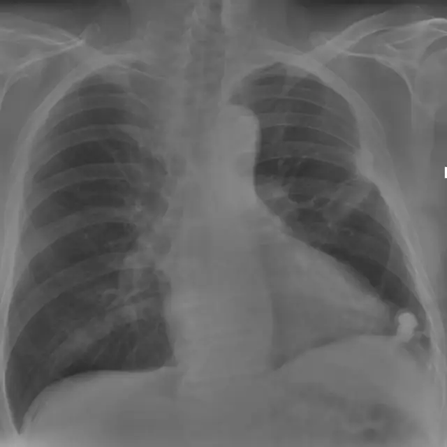
The silhouette sign: loss of a normal border means adjacent airspace disease. Air bronchograms mean the opacity is alveolar.
- 5
Hedging honestly
'Consistent with' (confident), 'suspicious for' (probable), 'cannot exclude' (possible but unproven), choose deliberately; clinicians act on your verbs.
Common mistakes
- Saying 'density' on MRI or 'echogenicity' on CT
- Using 'infiltrate' loosely, prefer 'consolidation' or 'opacity'
- Hedging every sentence until the report says nothing
Summary checklist
- Right term for the modality
- HU anchors memorized
- Sequence named on MRI
- Verbs chosen deliberately
Ask RADSpace about this guide
AI responses are for education and may contain errors. RADSpace is not for primary clinical diagnosis.