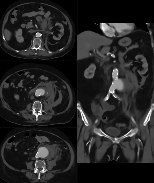
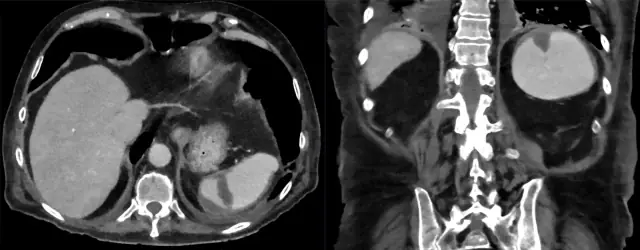
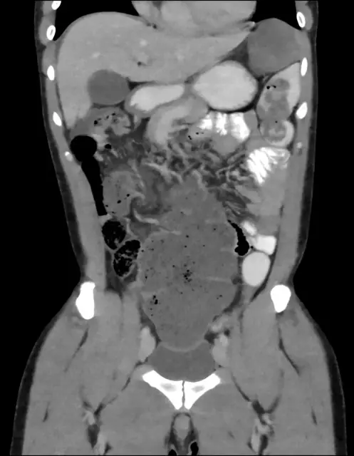
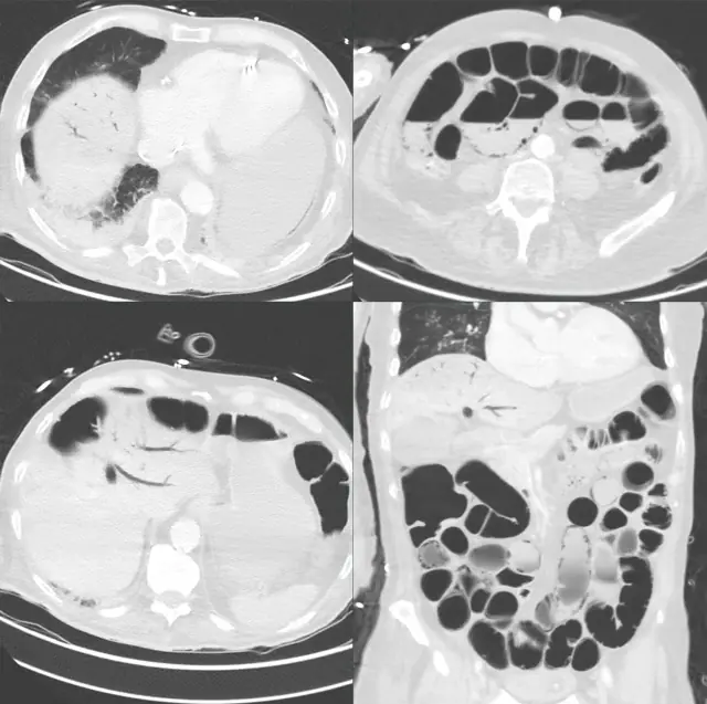
Recognizing free fluid on FAST
Learning objectives
- Know the four FAST windows and what each answers
- Identify free fluid confidently in Morison's pouch
- Understand FAST's decision role and limitations
Step-by-step approach
- 1
The question FAST answers
Is there free fluid (blood) in the peritoneal, pericardial, or pleural spaces of an unstable trauma patient? It is a decision tool, not a survey.
- 2
RUQ, Morison's pouch
The hepatorenal recess is the most dependent supine recess and the highest-yield single view. Free fluid = anechoic stripe between liver and kidney. Sweep the whole recess and the caudal liver tip.
- 3
LUQ
Fluid collects above the spleen (subphrenic) before the splenorenal recess, go a rib space higher and more posterior than you think.
- 4
Pelvis
The rectovesical or rectouterine pouch is the most dependent space overall; a full bladder is your acoustic window.
- 5
Pericardial
Subxiphoid view for pericardial fluid, see the pericardial effusion guide topics for tamponade signs.
- 6
Decision rule
Unstable + positive FAST → theatre. Stable → CT. Negative FAST does NOT exclude injury: it rules in, not out.
Common mistakes
- Declaring FAST negative from a single still image
- Missing subphrenic fluid in the LUQ by scanning too low
- Treating a negative FAST as excluding injury
Summary checklist
- Four windows
- Sweep each recess fully
- Liver tip checked
- Result stated per window
Ask RADSpace about this guide
AI responses are for education and may contain errors. RADSpace is not for primary clinical diagnosis.