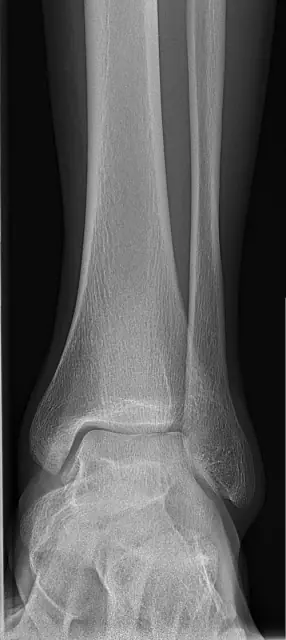
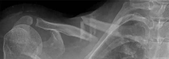
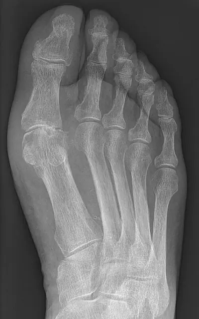
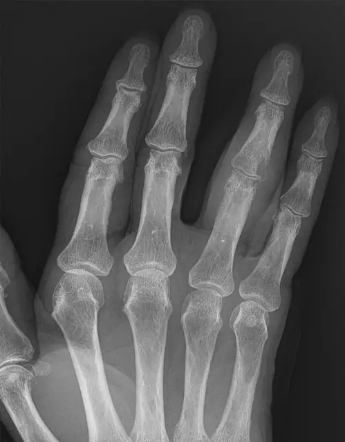
How to describe a fracture
Learning objectives
- Build a complete fracture description from six elements
- Communicate displacement and angulation unambiguously
- Know which features escalate urgency
Step-by-step approach
- 1
Bone and site
Name the bone and the segment: proximal/middle/distal third, metaphysis, diaphysis, or epiphysis; intra- vs extra-articular.
- 2
Pattern
Transverse, oblique, spiral, comminuted, segmental, avulsion, impacted, the pattern encodes the mechanism.
- 3
Displacement
Described by where the DISTAL fragment has gone, as a percentage of bone width where useful.
- 4
Angulation
Direction the apex points, or the tilt of the distal fragment, say which convention you're using. Dorsal angulation of the distal radius = Colles-type.
- 5
Joint involvement and special features
Intra-articular extension, dislocation, open fracture, and in children the physis (Salter-Harris).
- 6
The second fracture
Rings (pelvis, mandible, forearm) break in two places. Found one? Look for the other.
Common mistakes
- Vague descriptions ('broken wrist') that force the surgeon to re-read the film
- Missing the second fracture in ring structures
- Overlooking subtle impacted fractures, look for sclerotic bands
Summary checklist
- Bone + site
- Pattern
- Displacement
- Angulation
- Articular involvement
- Second fracture?
Ask RADSpace about this guide
AI responses are for education and may contain errors. RADSpace is not for primary clinical diagnosis.