How to approach a non-contrast CT head
Learning objectives
- Use the Blood–Cisterns–Brain–Ventricles–Bone sequence
- Recognize what acute blood looks like
- Understand windowing and why it matters
Step-by-step approach
- 1
Orient yourself
Axial slices are viewed from the feet: patient left is image right. Scroll the whole stack once quickly before detailed review.
- 2
Blood
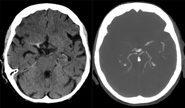
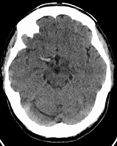
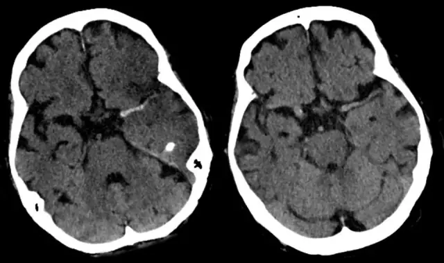
Acute blood is hyperdense (bright, ~50–70 HU). Check the parenchyma, the extra-axial spaces (subdural crescents, extradural lentiform collections), the sulci and cisterns (subarachnoid), and the ventricles.
- 3
Cisterns
Are the basal cisterns open? Effaced cisterns signal raised intracranial pressure and impending herniation.
- 4
Brain
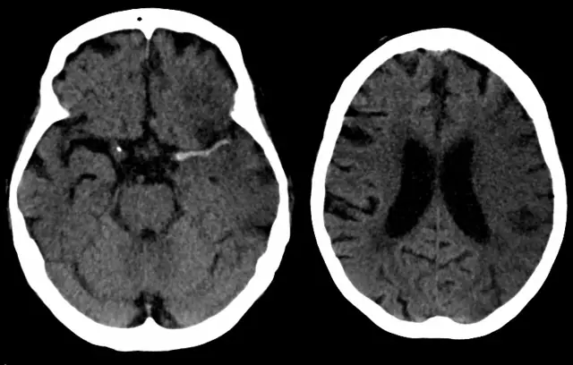
Grey–white differentiation preserved and symmetric? Loss of the insular ribbon or an obscured lentiform nucleus are early infarct signs.
- 5
Ventricles
Size and symmetry; midline shift; blood in dependent occipital horns.
- 6
Bone
Switch to bone windows. Fractures, especially where they cross vascular grooves, matter enormously.
Common mistakes
- Forgetting laterality, treating image left as patient left
- Reviewing only one window setting
- Missing subtle subdurals adjacent to bright skull, adjust windows
Summary checklist
- Blood
- Cisterns
- Brain
- Ventricles
- Bone
- Compare sides on every slice
Ask RADSpace about this guide
AI responses are for education and may contain errors. RADSpace is not for primary clinical diagnosis.