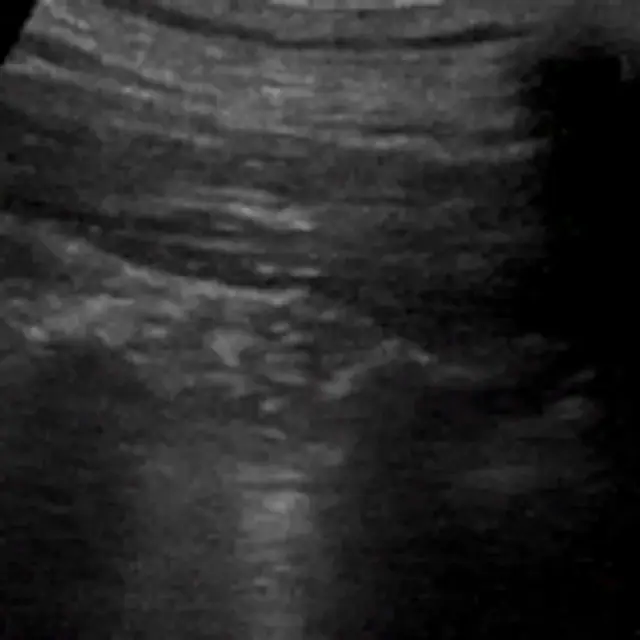
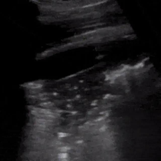
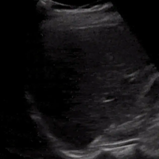
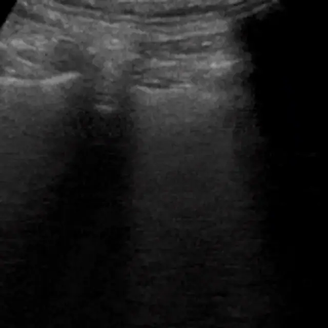
Basic cardiac POCUS views
Learning objectives
- Acquire the four core cardiac windows
- Answer the three big POCUS questions
- Recognize pericardial fluid and gross dysfunction
Step-by-step approach
- 1
The three questions
Basic cardiac POCUS answers: Is there a pericardial effusion? Is LV function grossly normal, reduced, or hyperdynamic? Is the RV dilated?
- 2
Parasternal long axis (PLAX)
Probe left of the sternum, 3rd–4th interspace, marker to the right shoulder. See LA, LV, aortic and mitral valves, RV outflow anteriorly.
- 3
Parasternal short axis
Rotate 90° from PLAX: the LV becomes a ring. A 'D-shaped' septum flattening toward the LV suggests RV pressure overload.
- 4
Apical four-chamber
At the apex beat, marker toward the bed (convention varies): all four chambers side by side. Compare RV:LV size, RV should be smaller.
- 5
Subxiphoid
Under the xiphoid using the liver as a window, often best in resuscitation and the classic view for effusion.
- 6
Eyeball assessment
Gross LV function is judged from endocardial excursion and wall thickening across several beats, trust the pattern across views, not one frame.
Common mistakes
- Judging function from a single beat or frame
- Confusing pericardial fat with effusion
- Over-reading RV size in an off-axis apical view
Summary checklist
- Effusion?
- LV function?
- RV size?
- At least two views obtained
Ask RADSpace about this guide
AI responses are for education and may contain errors. RADSpace is not for primary clinical diagnosis.